A Formidable Foe: Feline Odontoclastic Resorptive Lesions
 Cats have a habit of doing things their own way, and it is no different when it comes to dental disease. While they are subject to plaque, tartar, gingivitis, and periodontal disease just like their canine counterparts, they are also prone to a few conditions that are not quite so common in dogs.
Feline odontoclastic resorptive lesions are a progressive condition that we see in our cat patients at BEEVET Animal Hospital. By educating pet owners about this condition, we hope we can encourage more proactive dental health care for all of our patients, feline or not.
Recognizing Feline Odontoclastic Resorptive Lesions
The term feline odontoclastic resorptive lesions (FORLs) sounds daunting, but what exactly are they?
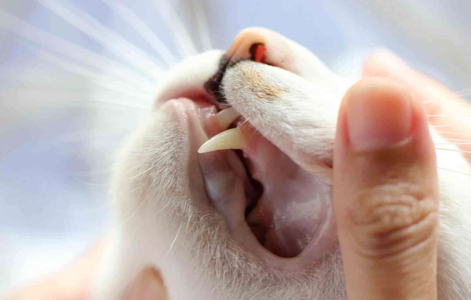
FORLs are a painful condition that results when cells within a normal tooth, called odontoblasts, begin to create holes within the tooth. These are typically near the gumline and are similar to a cavity, although their appearance happens for a different reason entirely.
As a lesion progresses, it can become infected due to the exposure of the tooth pulp. If extensive enough, the crown of the tooth may even fall off, leaving the roots intact below the gumline.
We don’t entirely understand what causes some cats to develop resorptive lesions, but we do know that over half of our feline patients over the age of 6 will be affected.
Cats who are suffering from this condition will often demonstrate signs of a problem as it advances, which may include:
- Drooling
- Bleeding from the mouth
- Reluctance to allow the mouth to be examined
- Difficulty eating
- Bad breath
In the early stages of this process, cats may not show obvious signs of an issue. Of course, we can often identify FORLs before they become troublesome with routine oral examinations and dental radiographs (x-rays).
Battling FORLs
Treatment for feline odontoclastic resorptive lesions depends a bit on the stage of the disease. This painful condition is not one to be ignored, however.
Once we have identified a FORL in a feline patient, we will assign a classification to the lesion:
Class I – A class one FORL is an early lesion in which the enamel covering of the tooth has a small defect noted. Treatment for these includes cleaning of the area and proactive home dental care, including fluoride.
Class II – A class two lesion is a little more serious, affecting not only the outer enamel but also the underlying dentin. In some situations, the enamel may be restored, but more often these teeth are extracted.
Class III – As a class two lesion progresses, it will eventually enter the pulp cavity of the tooth. These teeth should be extracted.
Class IV – When the crown of the tooth becomes fractured or eroded, the lesion is considered a class four. Gum tissue will often grow over the remaining pieces of root, creating the illusion that the tooth is entirely gone. These are often still quite painful.
Dental radiographs are essential for diagnosing and properly treating feline odontoclastic resorptive lesions. It is easy for these to hide from sight below the gums.
Unfortunately, because we don’t understand the cause for these lesions, it is impossible to fully prevent them. Often tooth extraction is the best course of action.
For now, providing your pet with routine examinations and proactive dental health care is the best we can do to battle feline odontoclastic resorptive lesions. Please give us a call with questions or to find out how we can help you provide this for your cat.